What Causes a plantar fibroma?
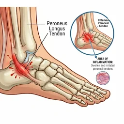
According to the National Library of Medicine, plantar fibromatosis, or plantar fibroma, is a non-cancerous condition characterized by the excessive growth of fibrous connective tissue in the superficial plantar fascia on the sole of the foot. Fascia is a fibrous tissue that helps to cover important blood vessels, nerves, tendons, and muscles. However unlike a tendon, fascia is not designed to allow for mobility and therefore is static. Therefore plantar fibromatosis, is an abnormal thickening of the foot’s deep connective tissue where nodules begin to grow in the fascia of the sole of the foot. We will talk about plantar fibroma treatment options, and how our staff can provide a positive outcome.
Plantar fibromatosis belongs to a family of diseases, Dupuytrens and Peyronie’s disease, where fundamentally the only difference is the location. Whereas Dupuytrens affects the hands and Peyronie’s male genitals. Plantar fibromatosis was first described in 1610 by George Ledderhose. These firm tender nodules presented within the central or the medial bands of the plantar fascia of the foot. Symptoms typically manifest gradually, and patients often seek medical attention once the condition progresses locally. This can result in discomfort, swelling, challenges with barefoot walking, and difficulties fitting into specific types of shoes.
- Plantar fibroma affects an estimated 200,000 people in the United States.
- It typically presents in middle-aged patients typically in the fourth and fifth decades of life.
- The disease is 2 times as likely to occur in males than females.
- It has an incidence of both feet in about 25% of patients.
There is a correlation with this disease being present in patients with a medical history of diabetes, epilepsy, or alcohol abuse disorder. This disease also has a strong familial inheritance pattern. Ledderhose disease shares a geographical similarity with Dupuytren, which is the area consistent with Northern European Nordic descent. It is hypothesized that this population which were originally considered “Vikings’ often had trauma holding onto weapons such as spears, swords, and hammers for an extensive period of time and with extreme blunt force.
Causes and Symptoms
A certain cell type called proliferating fibroblasts are thought to be the cause of plantar fascial nodules. These fibroblasts are located in areas with less cellular fascia and very little collagen and vascular supply.
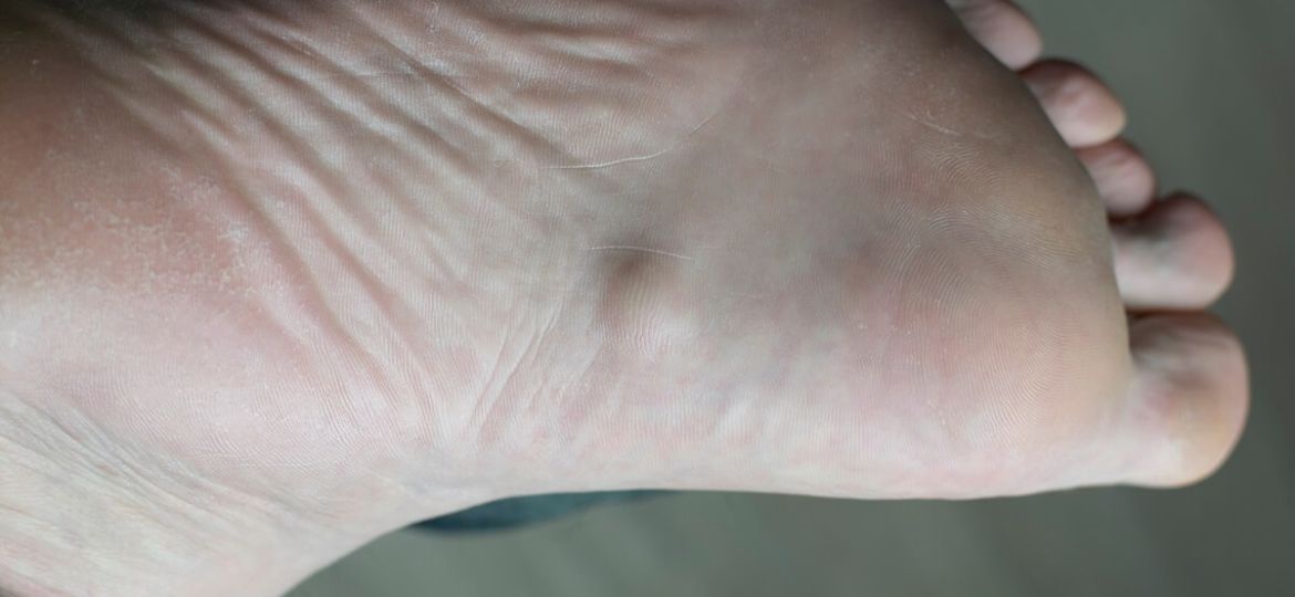
The classic presentation of plantar fibromatosis is an insidious slightly tender nodule in the medial or central band of the fascia of the foot that ranges anywhere from 0.5 to 3 cm in size. Other factors that should be taken into consideration when evaluating a plantar fibroma are any adjacent bony prominences which can be detected with an x-ray, tendon insertions, hindfoot alignment, as well as the presence of an Achilles or gastrocnemius Muscle contracture, which can exacerbate symptoms.
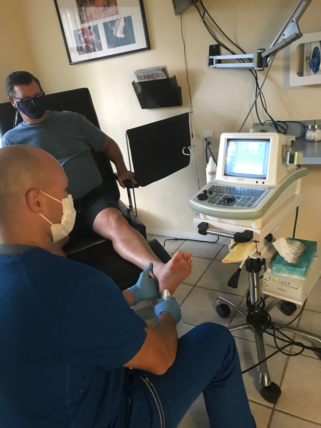
Aside from clinical evaluation, a combination of Ultrasound and MRI Foot Diagnosis at Certified Foot and Ankle Specialists can be used to exclude other potential causes including a lipoma, fibrosarcoma, or cyst. An ultrasound is useful to show the depth and size of the nodules.
The host of treatment options for plantar fibroma begin with conservative consist of stretching, dispensing of a night splint, orthotics and pads, steroid injections, and topical verapamil cream. Verapamil, commonly prescribed as a calcium channel blocker to manage hypertension, takes on a different role in treating plantar fibromatosis. In this context, it is employed to enhance collagenase activity while suppressing collagen production. One potential side effect of topical verapamil is contact dermatitis. For more intensive treatment, electron beam radiation therapy may be considered. This approach aims to reduce fibroblast activity by disrupting the production of transforming growth factor by fibroblasts. Consultation with a radiation oncologist would be appropriate should this be an avenue to be considered. Collagenase injections can also be applied into the site. Another option that is available in the St. Petersburg location is extracorporeal shockwave therapy. This tends to soften the nodes with time and leads to an overexpression of transforming growth factor beta causing an increased production of extracellular matrix components and restricting tissue contraction.
Treatment and Prevention of Plantar Fibroma
Surgical options are possible however, local incision has a recurrence rate from 60-100%, wide excision removal of the node with margins of 2 cm has a recurrence rate of up to 60%, and a complete fasciectomy with removal of the entire plantar fascia still has a recurrence rate of up to 25%. Because of the recurrence rates of failure, surgery should be considered a last resort.
Do you or a loved one suffer from what you believe may be a painful tender nodule on the bottom of the arch? If so, consult one of our Certified Foot and Ankle Specialists today to obtain more information and treatment options that work for you.
Dr Felipe Peterson, DPM