Introduction
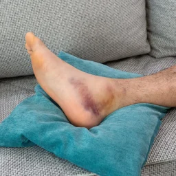
Foot pain is a prevalent complaint affecting millions of people worldwide, hindering mobility and diminishing quality of life. Diagnosing the root cause of foot pain accurately is crucial for effective treatment and improved patient outcomes. In recent years, medical technology has witnessed significant advancements in diagnostic imaging, with Magnetic Resonance Imaging (MRI) and the Pedcat CT Scanner emerging as invaluable tools in pinpointing the source of foot pain. This article explores how these advanced imaging techniques have revolutionized foot pain diagnosis, providing clinicians with unparalleled insights into complex foot pathologies.
Understanding Foot Pain Diagnosis from MRI
MRI has become a game-changer in the field of musculoskeletal imaging, offering detailed, high-resolution images of soft tissues, bones, and cartilage without the use of ionizing radiation. When it comes to diagnosing foot pain, MRI plays a vital role in identifying various conditions, such as:
A. Ligament and Tendon Injuries: MRI can reveal tears or degeneration in the ligaments and tendons, such as plantar fasciitis or Achilles tendonitis, which are common causes of foot pain.
B. Stress Fractures: Stress fractures, often challenging to detect on X-rays, become readily apparent on MRI scans, aiding clinicians in delivering early and precise interventions.
C. Foot Arthritis: MRI can detect early signs of arthritis, helping differentiate between different types of arthritis and guiding appropriate treatment strategies.
D. Nerve Compression: MRI is effective in identifying nerve compression syndromes, like Morton’s neuroma, allowing doctors to target the specific nerve for pain relief.
Pedcat CT Scanner: Unraveling Foot Pain in 3D
The Pedcat CT Scanner is an advanced imaging system specifically designed for musculoskeletal extremity imaging, including the foot and ankle. Unlike traditional CT scanners, the Pedcat focuses on delivering high-resolution 3D images with lower radiation exposure, making it safer and more suitable for specialized joint examinations.
A. Visualizing Bone Pathologies: The Pedcat CT Scanner provides exquisite detail of bone structures, enabling precise identification of stress fractures, bone tumors, and abnormalities that may contribute to foot pain.
B. Evaluating Joint Spaces: With its ability to capture multiple angles and planes, the Pedcat CT Scanner aids in assessing joint spaces for signs of arthritis, synovitis, or loose bodies, which are crucial factors in diagnosing foot pain.
C. Preoperative Planning: For patients requiring foot surgery, the Pedcat CT Scanner offers a comprehensive view of the affected area, empowering surgeons to plan and execute procedures with unparalleled accuracy.
Combining MRI and Pedcat CT: The Power of Fusion Imaging
While both MRI and the Pedcat CT Scanner excel in specific aspects of foot pain diagnosis, combining the two modalities through fusion imaging brings about a new level of diagnostic accuracy. Fusion imaging involves overlaying MRI and Pedcat CT data to create a comprehensive, multi-dimensional representation of the foot’s internal structures.
A. Improved Visualization: By combining the strengths of both techniques, fusion imaging provides a clearer and more detailed view of complex anatomical structures, facilitating the identification of subtle abnormalities.
B. Enhanced Diagnosis: The fusion of MRI and Pedcat CT data enables a more comprehensive assessment of the interaction between soft tissues and bones, leading to a more accurate diagnosis of conditions like osteochondral defects and cartilage injuries.
C. Personalized Treatment Plans: With fusion imaging, healthcare professionals can tailor treatment plans to the individual patient’s unique foot anatomy and pathology, resulting in improved outcomes and reduced recovery times.
Conclusion
In the quest to uncover the underlying causes of foot pain, modern medicine has embraced innovative diagnostic technologies like MRI and the Pedcat CT Scanner. These advanced imaging techniques have revolutionized foot pain diagnosis, empowering healthcare professionals to identify and treat various foot pathologies with unprecedented precision and accuracy. As technology continues to advance, we can expect even more refined imaging modalities, ushering in a new era of personalized medicine for individuals seeking relief from the burden of foot pain.
Certified Foot and Ankle Specialists was the first practice to offer patients a new, state-of-the-art option for diagnosing foot and ankle conditions: the pedCAT™. This compact machine generates standard X-rays and full CT scans in less than 60 seconds, reducing the time it takes to diagnose your condition and implement a treatment plan to get you back on your feet.
Find out more about the pedCAT™ and our practice here: