

Sarah Hubbard
*About the Author: Sarah Hubbard
Sarah Hubbard is a medical writer for Certified Foot and Ankle Specialists with approximately 15 years of experience creating accurate, patient-focused healthcare content. She specializes in translating complex podiatric topics into clear, informative articles that help patients better understand foot and ankle conditions, treatment options, and preventive care.
A navicular stress fracture is a small crack in the tarsal navicular bone in the midfoot, caused by repeated stress rather than a single injury.
This is considered a high-risk fracture because the center of the navicular has a poor blood supply, which slows healing and raises the chance of nonunion.
However, this is not the same as accessory navicular syndrome, which involves an extra bone some people are born with. Instead, a navicular stress fracture is a break in the normal bone caused by overuse. For instance, if you play tennis or pickleball across South Florida, this is one of the more common midfoot injuries we see in active adults over 40.
Fortunately, Certified Foot and Ankle Specialists is a multi-location podiatric surgery practice serving South Florida and Southwest Florida. Our board-certified foot and ankle surgeons diagnose and treat navicular stress fractures at convenient locations across Boca Raton, West Palm Beach, Fort Myers, and Cape Coral.
Quick Answers: Navicular Stress Fracture
- What is a navicular stress fracture? A hairline crack in the navicular bone of the midfoot from repetitive overload. It’s a high-risk fracture because of the bone’s limited blood supply.
- What are the symptoms? A vague ache on the top of the midfoot that gets worse with activity and eases with rest. Pinpoint tenderness over the top of the navicular, known as the N-spot, is a hallmark.
- How do doctors diagnose it? They use MRI or CT, not standard X-rays. Early X-rays look normal in most cases, which is why doctors often miss these fractures.
- Can it heal without surgery? Yes. Non-displaced fractures usually heal with 6–8 weeks of strict non-weight-bearing immobilization. Complete or displaced fractures may need surgery.
- How long is recovery? Most people return to full activity in 3–4 months. Surgical cases can take 4–6 months.
What is a navicular stress fracture?
A navicular stress fracture is a crack in the tarsal navicular, a boat-shaped bone that sits at the top of the midfoot arch. The navicular connects to the talus behind it and the three cuneiform bones in front, forming the keystone of the medial arch. Every step you take loads this bone, so repeated impact can create tiny cracks that don’t have time to heal between sessions.
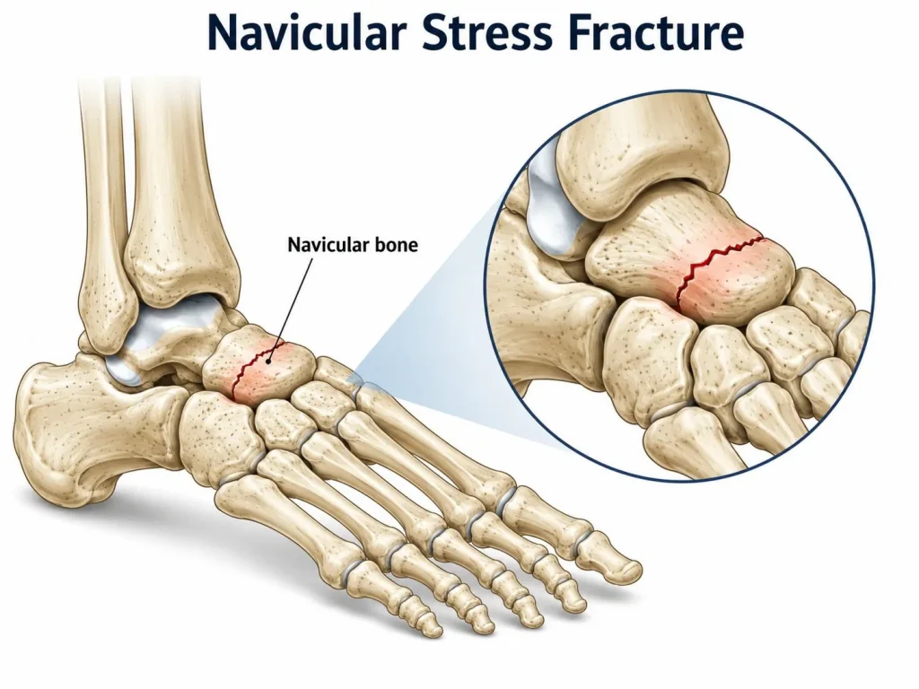 The reason this fracture worries foot and ankle surgeons is blood supply. The middle third of the navicular sits in a zone with naturally low circulation. According to the American Orthopaedic Foot & Ankle Society (AOFAS), this poor blood flow is why navicular stress fractures heal slowly and carry a real risk of nonunion, where the bone fails to knit back together.
What are the symptoms of a navicular stress fracture?
The main symptom is a dull, aching pain across the top of the midfoot that worsens with activity and fades with rest. Many patients describe it as a nagging ache they can’t quite locate, not a sharp injury. Because it builds slowly, people often keep training or playing for weeks before they get it checked.
Where does the pain show up?
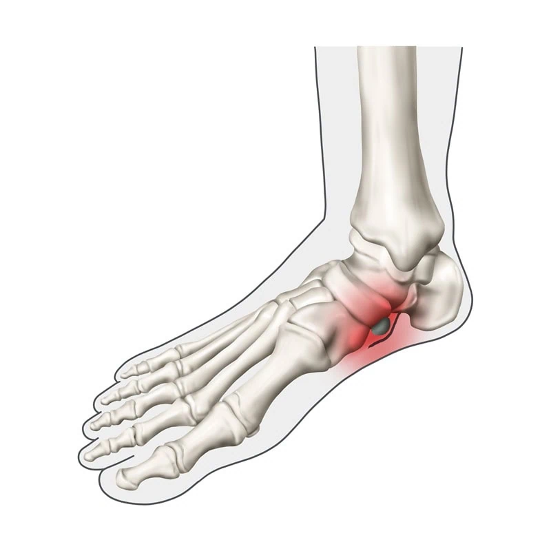
Pain and tenderness concentrate over the top of the navicular, a spot clinicians call the N-spot. Pressing directly on this area usually reproduces the pain. Some people also notice aching that spreads along the inner arch after long walks or a match.
How is it different from a sprain or tendonitis?
A sprain tends to follow a specific twist or roll, while a stress fracture builds gradually with no single trigger. Extensor tendonitis causes pain on top of the foot too, but it typically eases once you rest and warm up. Stress fracture pain returns every time you load the foot, which is the pattern that should prompt imaging.
What causes a navicular stress fracture?
Navicular stress fractures are caused by repetitive loading of the midfoot that outpaces the bone’s ability to repair itself. They show up most in runners, sprinters, basketball players, and dancers. In South Florida, we also see them in tennis and pickleball players, where quick stops and lateral cuts hammer the midfoot on hard courts.
Common contributing factors include:
- A sudden jump in training volume or court time without a gradual ramp-up
- Hard playing surfaces that increase repetitive impact
- A high-arched (cavus) foot that concentrates load on the midfoot
- Tight calf muscles that shift force forward into the arch
- Worn-out or unsupportive footwear
None of these cause a systemic problem on their own. A stress fracture is a mechanical overload injury, and identifying the load pattern behind it is part of preventing the next one.
Why is a navicular stress fracture considered high-risk?
A navicular stress fracture is high-risk because the central third of the bone has a limited blood supply, which slows healing and can lead to nonunion. Bones need steady blood flow to deliver the cells that rebuild them. The middle of the navicular sits in a watershed zone where two blood supplies meet and neither reaches strongly, so a crack there mends far slower than a fracture in a well-supplied bone.
Left untreated, these fractures can progress to a complete break, delayed union, or in some cases avascular necrosis, where part of the bone dies from lack of blood flow. A 2003 review in American Family Physician noted that navicular stress fractures 1 are frequently missed on first presentation, which delays treatment and worsens outcomes. That’s why early diagnosis and strict non-weight-bearing matter so much with this specific bone.
How is a navicular stress fracture diagnosed?
A navicular stress fracture is diagnosed with MRI or CT, because standard X-rays usually look normal in the early stages. This is the single biggest reason these injuries get missed. A patient gets an X-ray, it reads clean, and they’re told to rest, while the fracture keeps progressing.
A thorough workup usually follows this order:
- Clinical exam — the specialist checks for focal tenderness over the N-spot and pain with hopping or single-leg loading.
- MRI — detects the stress reaction and early fracture line before it shows on X-ray.
- CT scan — the standard for confirming how complete the fracture is and guiding treatment.
If you’ve had midfoot pain for more than two weeks and an X-ray came back normal, that doesn’t rule out a stress fracture. It’s a reason to ask a foot and ankle specialist about advanced imaging.
How long does a navicular stress fracture take to heal?
Most navicular stress fractures need 6–8 weeks of strict non-weight-bearing immobilization, with a total recovery of 3–4 months before full activity. Strict means no weight on the foot at all, usually in a cast or boot with crutches or a knee scooter. Partial weight-bearing during this window is one of the main reasons these fractures fail to heal.
Whether you need surgery depends on the fracture. The table below compares the two main paths, and your specialist decides based on your CT findings and activity demands.
| Factor | Conservative (non-weight-bearing cast) | Surgical (ORIF screw fixation) |
|---|---|---|
| Best for | Early or incomplete fractures caught before displacement | Complete fractures, displaced fractures, or nonunion |
| Incision required | No | Yes |
| Weight-bearing | None for 6–8 weeks | Protected, often earlier return under guidance |
| Hardware | None | One or two compression screws |
| Typical recovery | 3–4 months to full activity | 4–6 months, faster for high-demand athletes |
| When used at CFAS | First-line for most non-displaced fractures | When imaging shows a complete break or healing stalls |
Do you need surgery for a navicular stress fracture?
Patients require surgery when the fracture is complete or displaced, when healing stalls, or when high-demand athletes need a reliable return to sport. The procedure is an open reduction and internal fixation (ORIF), where a surgeon secures the bone with one or two compression screws. According to the American College of Foot and Ankle Surgeons (ACFAS), screw fixation of complete navicular stress fractures produces high union rates and a more predictable recovery than continued conservative care once a fracture has fully formed.
For a non-displaced fracture caught early, though, casting and non-weight-bearing usually work well and avoid surgery altogether. The right call depends on imaging, not guesswork.
When should you see a foot and ankle specialist?
See a foot and ankle specialist if midfoot pain lasts more than two weeks, returns every time you resume activity, or makes it hard to bear weight. Catching a navicular stress fracture early is the difference between a cast and an operation. These are red flags worth acting on:
- Aching on the top of the midfoot that keeps coming back after rest
- Pain that flares every time you return to running or the court
- Tenderness when you press on the top of the arch (the N-spot)
- A normal X-ray but pain that won’t settle
- Trouble putting full weight through the foot
The bottom line
A navicular stress fracture is a slow-healing midfoot injury that rewards early diagnosis and punishes delay. Most cases heal without surgery when they’re caught early and treated with strict non-weight-bearing, but a missed fracture can turn into a much harder problem. If you’re dealing with stubborn midfoot pain in South Florida request an appointment with one of our podiatrists. Our foot and ankle surgeons can confirm the diagnosis with advanced imaging and build the right plan, whether that’s conservative care or surgical fixation.
References:
- American Family Physician. 2003;67(1):85-91. Tarsal Navicular Stress Fractures
⚠️ MEDICAL DISCLAIMER
Timeframes, protocols, and clinical indications may vary.
Individual treatment outcomes differ significantly based on patient age, bone quality, fracture complexity, overall health, and compliance. This table is for educational purposes only and does not replace professional medical consultation.
Consult your orthopedic physician or qualified healthcare provider before making any treatment decisions. Treatment plans must be personalized based on your specific medical condition, imaging findings, and individual circumstances.
Frequently Asked Questions: Accessory Stress Fracture
Q: What does a navicular stress fracture feel like?
A: A navicular stress fracture usually feels like a dull ache across the top of the midfoot that worsens with activity and eases with rest. The pain builds gradually rather than starting with a single injury, and it often returns each time you resume running or court sports.
Q: Does accessory navicular syndrome go away on its own?
A: Early navicular stress fractures often look normal on standard X-rays because the crack is too fine to see. MRI or CT is needed to confirm the fracture, which is why these injuries are frequently missed at first and require advanced imaging when midfoot pain persists.
Q: Can a navicular stress fracture heal without surgery?
A: Yes. Most non-displaced navicular stress fractures heal with 6 to 8 weeks of strict non-weight-bearing immobilization in a cast or boot, followed by a gradual return to activity. Complete or displaced fractures, or fractures that fail to heal, may need surgical fixation.
Q: How long am I non-weight-bearing with a navicular stress fracture?
A: Strict non-weight-bearing typically lasts 6 to 8 weeks for a navicular stress fracture. Putting partial weight on the foot too early is a common reason these fractures fail to heal, so specialists usually require crutches, a knee scooter, or a cast during this period.
Q: What happens if a navicular stress fracture goes untreated?
A: An untreated navicular stress fracture can progress to a complete break, delayed union, or nonunion, and in some cases avascular necrosis where part of the bone dies from poor blood supply. Early diagnosis and strict non-weight-bearing greatly improve the outcome.