

Sarah Hubbard
*About the Author: Sarah Hubbard
Sarah Hubbard is a medical writer for Certified Foot and Ankle Specialists with approximately 15 years of experience creating accurate, patient-focused healthcare content. She specializes in translating complex podiatric topics into clear, informative articles that help patients better understand foot and ankle conditions, treatment options, and preventive care.
A sesamoid fracture is a break in one of the two small bones embedded beneath the first metatarsal head, at the base of the big toe, caused either by a direct impact or by the cumulative stress of repetitive forefoot loading.
Sesamoid fractures are among the most commonly misdiagnosed forefoot injuries. Because the pain closely resembles sesamoiditis and turf toe, patients frequently go weeks without accurate imaging — and delay in diagnosis is the single biggest predictor of prolonged recovery or non-union. If you suspect a sesamoid fracture, early evaluation and the right imaging protocol make a significant difference in outcome.
Certified Foot and Ankle Specialists is a multi-location podiatric surgery practice serving South Florida and Southwest Florida, with board-certified foot and ankle surgeons diagnosing and treating sesamoid fractures at locations across Boca Raton, Delray Beach, West Palm Beach, Fort Myers, and Cape Coral.
Quick Answers: Sesamoid Fracture
Can you walk on a sesamoid fracture? Often yes, but with significant pain. Many patients walk on an undiagnosed sesamoid fracture for days or weeks. Walking doesn’t confirm or rule out a fracture, X-ray and MRI do. Continued weight-bearing without treatment can progress a stress fracture to complete fracture or avascular necrosis.
What are the symptoms of a sesamoid fracture? Sharp or aching pain beneath the big toe joint that worsens with push-off and activity. Bruising, swelling, and tenderness directly beneath the first metatarsal head are typical. Acute fractures cause sudden severe pain after impact; stress fractures build gradually over weeks.
How is a sesamoid fracture treated? First-line treatment is non-surgical: a walking boot for 6-8 weeks combined with sesamoid offloading padding and activity restriction. Stress fractures may require 10-16 weeks of immobilization. Ssesamoidectomy surgery is reserved for non-union cases that don’t respond to 3-6 months of conservative care.
How long does a sesamoid fracture take to heal? Acute fractures typically heal in 3-4 months with appropriate immobilization. Stress fractures take longer, often 4-6 months, because of the sesamoid’s limited blood supply. Complicated cases involving avascular necrosis or non-union can extend to 12 months or require surgery.
When is sesamoid fracture surgery needed? Surgery is considered when 3-6 months of conservative treatment — boot immobilization, orthotics, and activity modification has not produced adequate healing. Sesamoidectomy (removal of the fractured sesamoid) carries a high success rate for appropriate candidates but requires careful planning to avoid toe deformity.
What is a sesamoid fracture, and how is it different from sesamoiditis?
A sesamoid fracture is a structural break in one or both of the sesamoid bones, a distinctly different injury from sesamoiditis, which is inflammation of the sesamoid and surrounding tendon without a fracture line.
The distinction matters clinically because treatment, immobilization duration, and return-to-activity timelines differ significantly between the two conditions. Sesamoiditis typically responds to orthotics and activity modification within 4-8 weeks. A sesamoid fracture requires 3-6 months of structured immobilization and carries a risk of non-union if undertreated.
A third diagnostic consideration is the bipartite sesamoid, a normal anatomic variant where the medial sesamoid forms in two pieces during development. Bipartite sesamoids appear in roughly 10-30% of people and are frequently misread as fractures on X-ray. The edges of a bipartite sesamoid are smooth and rounded; fracture edges are irregular and jagged. MRI and comparison X-rays of the opposite foot are the most reliable tools for distinguishing the two.
For a deeper look at sesamoid bone anatomy and how sesamoiditis differs from fracture, see our sesamoid bone anatomy guide.
What are the symptoms of a sesamoid fracture?
The hallmark of an acute sesamoid fracture is sudden, sharp pain beneath the first metatarsal head immediately following an impact such as a jump landing, a hard step, or a direct blow to the forefoot.
Stress fractures develop differently: pain builds gradually over several weeks of repetitive activity, often without a single identifiable incident. This pattern is common in runners, dancers, and athletes who sharply increase training volume without adequate progression.
Symptoms that distinguish a sesamoid fracture from sesamoiditis or turf toe:
- Pain localized directly beneath the first metatarsal head, not along the toe joint
- Bruising on the plantar surface of the foot is often absent with sesamoiditis
- Swelling that doesn’t resolve with a few days of rest and ice
- Worsening pain with push-off, stair climbing, or walking on hard surfaces
- Inability to fully extend the big toe without significant pain
Stress fracture symptoms tend to be more diffuse and are often described as a deep ache rather than sharp pain. Many patients are surprised by a fracture diagnosis because the pain never felt severe enough to suggest a broken bone.
How do podiatrists diagnose a sesamoid fracture?
Sesamoid fracture diagnosis requires a structured imaging protocol. A physical exam alone cannot distinguish a fracture from sesamoiditis or a bipartite sesamoid.
At Certified Foot and Ankle Specialists, diagnosis begins with a focused history of onset, activity level, and footwear, followed by a physical exam that includes direct palpation of both sesamoid bones and a plantarflexion stress test of the first MTP joint.
Imaging options and when each is used
- Weight-bearing X-rays (standard views + sesamoid view) – first-line imaging. Rules out obvious fracture and identifies bipartite sesamoid. The sesamoid-axial view is essential; standard AP and lateral views frequently miss sesamoid pathology.
- MRI – preferred when X-rays are inconclusive. The only imaging modality that reliably differentiates sesamoiditis from stress fracture and identifies avascular necrosis. Bone marrow edema on T2-weighted MRI is the diagnostic marker for acute stress fracture.
- PedCAT weight-bearing CT scan – available at CFAS for complex cases. Provides 3D anatomy under load, particularly useful when standard X-ray views give ambiguous results for bipartite sesamoid anatomy.
- Bone scan – used when MRI is not available or contraindicated; less specific than MRI for identifying fracture type.
A key clinical note: comparison X-rays of the opposite foot are valuable for distinguishing bipartite sesamoid from acute fracture. If the contralateral foot shows the same two-piece sesamoid, the finding is anatomic not traumatic.
How is a sesamoid fracture treated without surgery?
Conservative treatment resolves most sesamoid fractures when immobilization is adequate and maintained for the full required period. Early termination of the boot phase is the most common reason for non-union.
Acute sesamoid fracture protocol
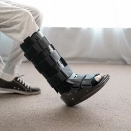
- Boot immobilization – A below-knee walking boot or stiff-soled post-op shoe enforces non-weight-bearing or partial weight-bearing for 6-8 weeks. The goal is complete elimination of push-off forces across the fracture site.
- Sesamoid offloading – Sesamoid padding (dancer’s pad or J-pad) positioned to offload the fractured sesamoid within the boot or post-op shoe. Continues through the transition out of the boot.
- Activity restriction – No running, jumping, dancing, or sport-specific loading. Swimming and upper-body training are permitted. Low-impact cycling (with flat pedals, not cleats) is typically allowed after 2-3 weeks.
- Anti-inflammatory management – NSAIDs for the first 1-2 weeks. Some evidence suggests prolonged NSAID use may impair bone healing — your CFAS podiatrist will guide appropriate duration.
- Custom orthotics at transition – Custom orthotic with sesamoid cut-out replaces boot at 6-8 weeks. Worn for a further 4-6 weeks during graduated return to activity.
Stress fracture protocol (extended timeline)
Stress fractures have a longer immobilization requirement, typically 10-16 weeks in a boot, because they represent cumulative bone damage rather than a single traumatic event. Returning to activity too early is the primary cause of stress fracture non-union.
- Bone stimulation – low-intensity pulsed ultrasound (LIPUS) devices accelerate bone healing in cases of delayed union. CFAS prescribes these for stress fractures that show incomplete healing at the 8-week imaging checkpoint.
- Calcium and vitamin D supplementation – checked and addressed if deficient, particularly in female runners and dancers with high stress fracture risk profiles.
- EPAT (shockwave therapy) – available at CFAS for cases of delayed healing or early non-union where bone stimulators have not produced adequate response. Stimulates vascularization and bone remodeling in the fracture zone.
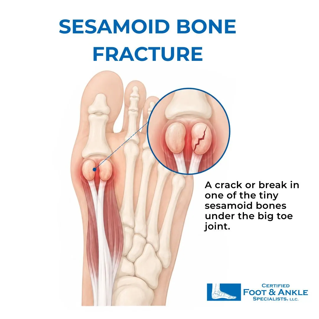
Sesamoid Fracture Treatment: Conservative vs. Surgical
| Factor | Conservative (Boot) | Intermediate (EPAT / Bone Stimulator) | Sesamoidectomy (Surgery) |
|---|---|---|---|
| Best for | Acute fractures + most stress fractures | Delayed union at 8-12 week imaging check | Non-union after 3-6 months documented conservative care |
| Duration | 6-16 weeks boot + 4-6 weeks orthotics | 6-12 weeks alongside orthotics | Surgery + 3-4 months post-op |
| Weight-bearing | Partial (boot) → full (orthotics) | Full in boot during treatment | Non-weight-bearing 2-3 weeks post-op |
| Return to sport | 12-20 weeks from diagnosis | 16-24 weeks | 4-6 months post-op |
| Success rate | 85-90% for acute, 70-80% for stress fracture | 70-80% for delayed union cases | Good for appropriate candidates, risk of hallux drift if medial sesamoid removed |
| When used at CFAS | All presentations — first-line | At 12 week review if healing incomplete | After 3-6 months failed conservative treatment, or avascular necrosis confirmed on MRI |
How long does a fractured sesamoid bone take to heal?
Sesamoid fractures heal slowly because both sesamoid bones have a limited, end-arterial blood supply — there are no collateral vessels to compensate if the primary blood supply is compromised. This is why immobilization duration is non-negotiable.
| Fracture/Condition Type | Boot Duration | Total Recovery | Return to Sport | Complication Risk |
|---|---|---|---|---|
| Acute sesamoid fracture | 6-8 weeks | 3-4 months | 12-16 weeks | Low if treated promptly |
| Sesamoid stress fracture | 10-16 weeks | 4-6 months | 16-20 weeks | Moderate — higher non-union rate |
| Non-union (failed healing) | N/A — reassess treatment | 6-12 months | Case-by-case | High — may require EPAT or surgery |
| Avascular necrosis | N/A — surgical evaluation | 6-12+ months post-op | 4-6 months post-op | High — bone death requires sesamoidectomy |
According to the American College of Foot and Ankle Surgeons (ACFAS), sesamoid fractures that receive adequate early immobilization have an 85-90% rate of complete union. Those that are undertreated or return to activity prematurely have significantly higher rates of non-union requiring surgical intervention.
Imaging at 6-8 weeks (X-ray) and 12 weeks (MRI if healing is incomplete) allows your CFAS podiatrist to assess whether the fracture is consolidating on schedule and adjust the protocol before non-union becomes established.
When does a sesamoid fracture require surgery?
Sesamoid fracture surgery, called sesamoidectomy, is indicated when 3-6 months of documented conservative care has not produced adequate fracture healing.
The procedure involves surgical excision of the fractured sesamoid through a small incision on the plantar or dorsal surface of the foot. Medial sesamoidectomy carries a specific risk: removal of the medial sesamoid can destabilize the medial plantar support of the first MTP joint, leading to hallux valgus (bunion drift) in susceptible patients. This risk is higher in patients with pre-existing pronation or a low first metatarsal declination angle. Lateral sesamoidectomy is generally less complex with fewer secondary deformity risks.
Two additional indications for earlier surgical consideration:
- Avascular necrosis of the sesamoid – confirmed on MRI when T1 signal shows bone death. Conservative treatment rarely reverses established AVN; surgical intervention is typically indicated once the diagnosis is made.
- Displaced fracture – when imaging shows significant fragment displacement, conservative immobilization may not bring the fragments into contact for healing. Surgical fixation or excision is evaluated on a case-by-case basis.
At CFAS, surgical decisions for sesamoid fracture are made after a thorough review of serial imaging, biomechanical profile, and documented conservative care history. Our board-certified podiatric surgeons discuss every surgical option, including the risks specific to medial vs. lateral sesamoidectomy, before proceeding.
When should you see a podiatrist for ball of foot pain?
See a foot specialist within 5-7 days if you have pain beneath the first metatarsal head that doesn’t improve with rest, ice, and reduced activity.
Don’t wait if:
- Pain was triggered by a specific impact such as a jump landing, a hard step, or a direct blow
- You’re an athlete or dancer who has recently increased training volume significantly
- Swelling or bruising appeared beneath the ball of the foot
- Pain is worsening rather than improving after 48-72 hours of rest
- You have diabetes, osteoporosis, or a history of prior foot fractures
Early diagnosis and accurate imaging prevent the most common complication of sesamoid fractures: delayed treatment that allows a stress fracture to progress to non-union. A fracture caught at 2-3 weeks responds very differently to treatment than one identified at 2-3 months.
If you’ve been treating forefoot pain as sesamoiditis and it’s not improving, ask for imaging that specifically includes the sesamoid-axial X-ray view. Standard foot X-rays frequently miss sesamoid pathology.
For patients dealing with sesamoiditis specifically rather than a fracture, our sesamoiditis treatment guide covers that condition in full.
Get Evaluated At Certified Foot And Ankle Specialists For A FractureD Sesamoid Bone
Sesamoid fractures heal well when caught early and immobilized properly. The key is not letting a gradual-onset injury go undiagnosed for months.
If you’re experiencing pain beneath the ball of your foot in Boca Raton, Delray Beach, West Palm Beach, Boynton Beach, or Coral Springs, Dr. Ashley Bowles, DPM, FACFAS, CWSP can evaluate you with same-visit X-rays and build an immobilization plan calibrated to your fracture type and activity level. For patients in Fort Myers or Cape Coral, Dr. Victor L. Herrera, DPM, ABMSP, FPMA, APMA leads our Southwest Florida sesamoid fracture evaluations.
Contact Certified Foot and Ankle Specialists to schedule your evaluation. Same-visit X-rays, MRI referrals, and in-office PedCAT scanning available at select locations.
Frequently Asked Questions Frequently Asked Questions About Sesamoid Bone Fracture
Q: Can you walk on a sesamoid fracture?
A: Yes, many patients walk on an undiagnosed sesamoid fracture for days or weeks with manageable pain. Walking doesn’t confirm or rule out a fracture. Continued weight-bearing without a boot or offloading increases the risk of non-union or progression from stress fracture to complete fracture. X-ray and MRI are required for accurate diagnosis before determining weight-bearing status.
Q: How long do you wear a boot for a sesamoid fracture?
A: Acute sesamoid fractures typically require 6-8 weeks in a walking boot followed by 4-6 weeks in custom orthotics with sesamoid offloading — a total of 3-4 months before return to full activity. Stress fractures require a longer boot phase of 10-16 weeks due to slower healing from the sesamoid’s limited blood supply.
Q: What is the difference between a sesamoid fracture and a bipartite sesamoid?
A: A bipartite sesamoid is a normal anatomic variant where the medial sesamoid forms in two pieces during development — present in roughly 10-30% of people. It is not a fracture and does not require treatment. The key imaging distinction is edge morphology: bipartite sesamoid edges are smooth and rounded; fracture edges are irregular and jagged. Comparison X-rays of the opposite foot and MRI are the most reliable tools for distinguishing the two.
Q: How long does a sesamoid fracture take to heal?
A: Acute sesamoid fractures heal in approximately 3-4 months with adequate boot immobilization and offloading. Stress fractures take 4-6 months due to the sesamoid’s limited blood supply. Cases complicated by delayed diagnosis, early return to activity, or avascular necrosis can extend to 12 months or require surgical intervention.
Q: What happens if a sesamoid fracture doesn’t heal?
A: A sesamoid fracture that fails to heal after adequate conservative treatment becomes a non-union — two fracture fragments that remain separated without bridging bone. Non-union causes persistent forefoot pain with activity. It is treated with either extended immobilization combined with EPAT (shockwave therapy) or bone stimulation, or surgical removal of the affected sesamoid (sesamoidectomy). Avascular necrosis — bone death from compromised blood supply — is a more serious complication requiring earlier surgical consideration.
Q: Is sesamoid bone surgery serious?
A: Sesamoid bone surgery (sesamoidectomy) is an outpatient procedure with a high success rate for appropriate candidates. The main risk specific to medial sesamoidectomy is postoperative hallux valgus (bunion drift), which can occur when the medial plantar support of the first MTP joint is destabilized. Lateral sesamoidectomy carries fewer secondary deformity risks. Most patients return to full activity within 4-6 months post-surgery.
Q: Can a sesamoid fracture heal without surgery?
A: Yes, 85-90% of acute sesamoid fractures heal without surgery when boot immobilization and offloading are maintained for the full required duration (6-8 weeks for acute fractures, 10-16 weeks for stress fractures). Surgery is reserved for cases where 3-6 months of documented conservative treatment has failed to produce healing, or when avascular necrosis is confirmed on MRI.